


pCD2-hTL1A-Tg
Nomenclature
C57BL/6JSmo-Tgtn(pCD2-hTL1A)1Smoc
Cat. NO.
NM-TG-250279
Strain State
Sperm cryopreservation
Model Description
Validation Data

Fig.1 Detection of human TL1A expression in serum by ELISA (n=3, 6 weeks old).
Abbr. WT, wild type; N.D. not detected.
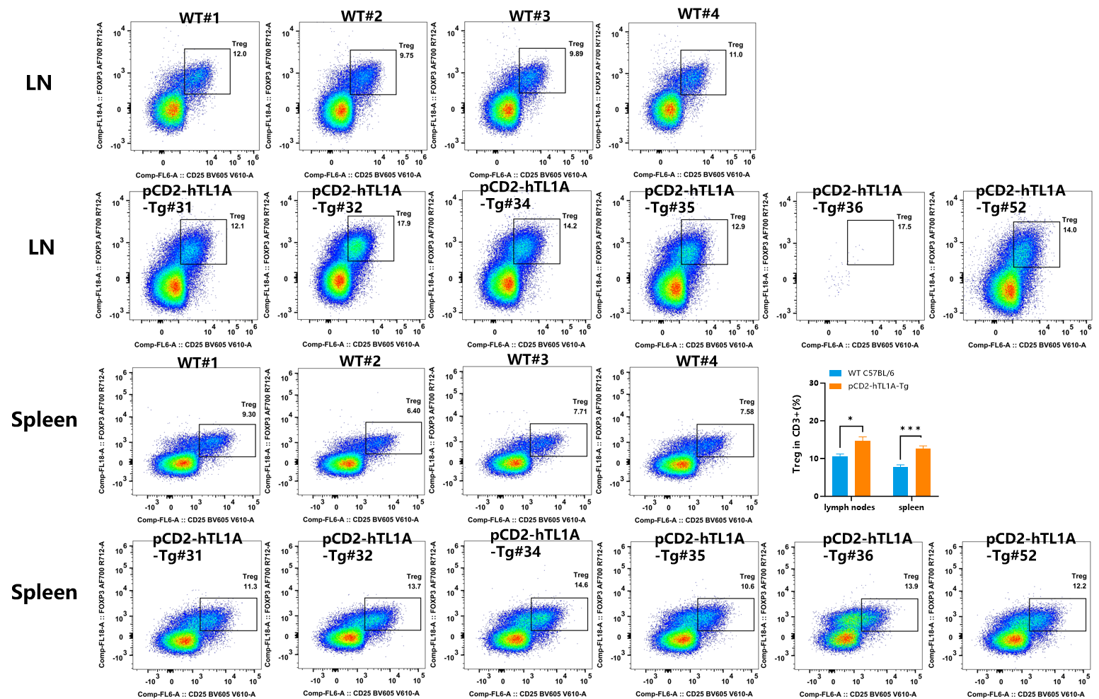
Fig.2 Detection of Treg expression on CD3+T cells in MLN and spleen of WT (C57BL/6) mice and pCD2-hTL1A-Tg mice by FACS.
In CD3+ T cells, compared with wild-type mice, the number of Treg cells in the spleen and MLN of pCD2-hTL1A-Tg mice was increased (n=4-6, 17 weeks old).
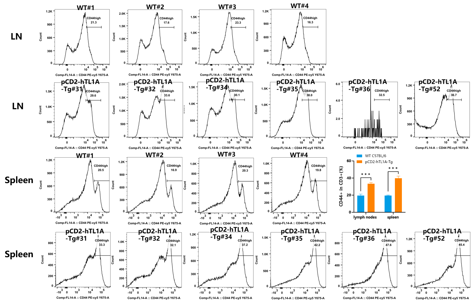
Fig.3 Detection of CD44 expression on CD3+T cells in MLN and spleen of WT (C57BL/6) mice and pCD2-hTL1A-Tg mice by FACS.
In CD3+ T cells, compared with wild-type mice, the number of CD44+ cells in the spleen and MLN of pCD2-hTL1A-Tg mice was increased (n=4-6, 17 weeks old).
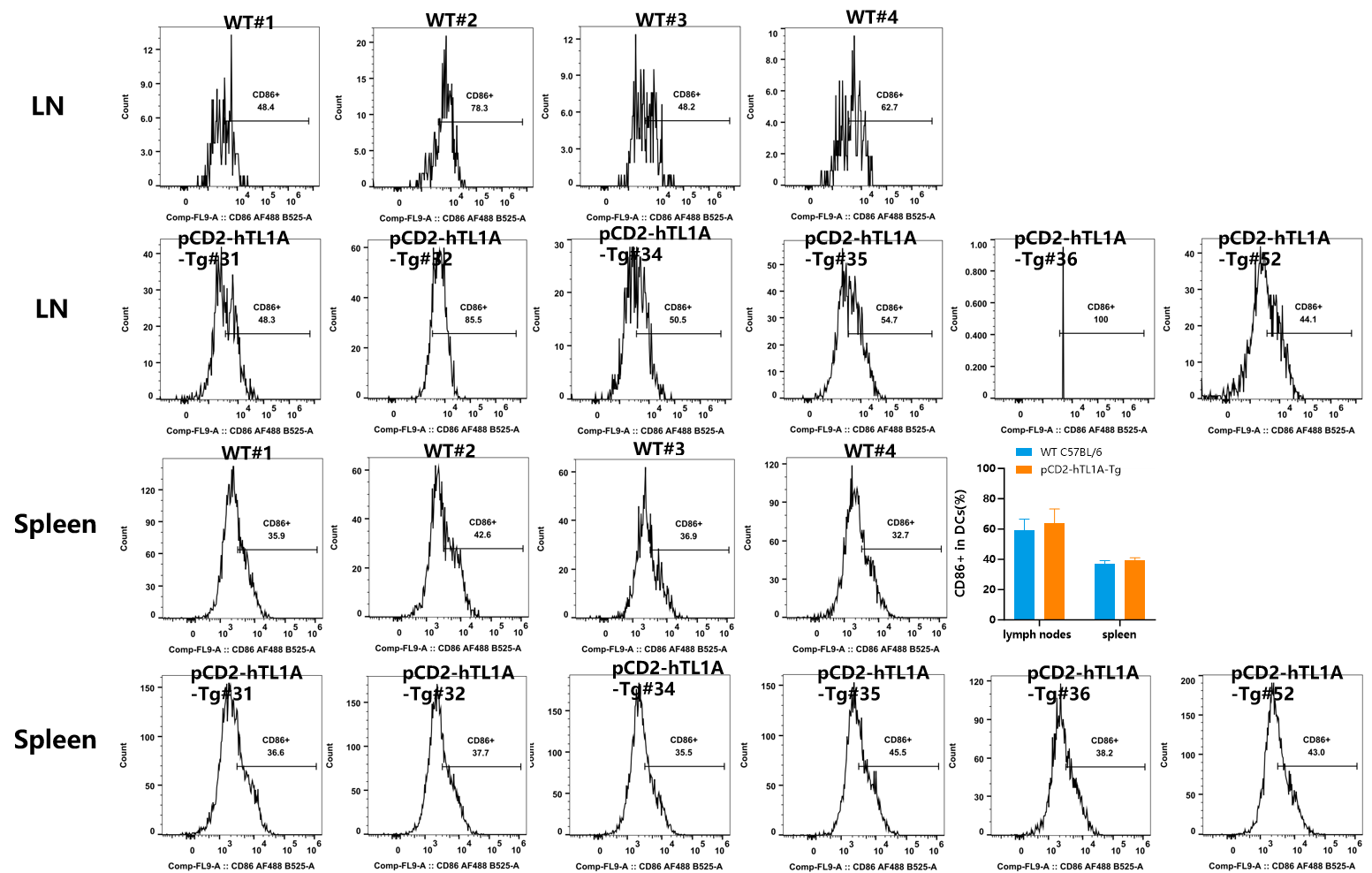
Fig.4 Detection of CD86 expression on CD11c+DCs in MLN and spleen of WT (C57BL/6) mice and pCD2-hTL1A-Tg mice by FACS.
In DCs cells, the number of CD86+ cells in the spleen and MLN of pCD2-hTL1A-Tg mice was similar to that of wild-type mice (n=4-6, 17 weeks old).
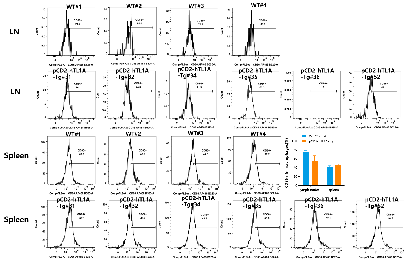
Fig.5 Detection of CD44 expression on macrophages in MLN and spleen of WT (C57BL/6) mice and pCD2-hTL1A-Tg mice by FACS(n=4-6, 17 weeks old).
In macrophages, the number of CD86+ cells in the spleen and MLN of pCD2-hTL1A-Tg mice was similar to that of wild-type mice.


Fig.6 Detection of IFN-γ expression on CD4+T cells in MLN and spleen of WT (C57BL/6) mice and pCD2-hTL1A-Tg mice by FACS.
In CD4+ T cells, compared with wild-type mice, the number of IFNγ+ cells in the spleen and MLN of pCD2-hTL1A-Tg mice was significantly increased (n=4-6, 17 weeks old).

Fig.7 Detection of IL-17A+ expression on CD4+T cells in MLN and spleen of WT (C57BL/6) mice and pCD2-hTL1A-Tg mice by FACS.
In CD4+ T cells, compared with wild-type mice, the number of IL-17A+ cells in the spleen and MLN of pCD2-hTL1A-Tg mice was significantly increased (n=4-6, 17 weeks old).

Fig.8 Detection of IL-4+ expression on CD4+T cells in MLN and spleen of WT (C57BL/6) mice and pCD2-hTL1A-Tg mice by FACS.
No IL-4+ cells were observed in the spleen and MLN of pCD2-hTL1A-Tg mice and wild-type mice (n=4-6, 17 weeks old).

Fig. 8 Phenotype of WT (C57BL/6) mice and pCD2-hTL1A-Tg mice (n=2-3, 17 weeks old). (A) Gross photograph; (B) Colon photo; (C) Body weight and intestine index.

Fig. 9 H&E staining of the small intestine and colon in pCD2-hTL1A-Tg mice (n=2-3, 17 weeks old).

Fig. 10 Representative masson's trichrome staining images of the small intestine and colon in pCD2-hTL1A-Tg mice. (n=2-3, 17 weeks old)
You may also like
Shanghai Model Organisms Center Inc has licensed CRISPR-Cas9 technology from Broad Institute
On Dec 16, 2018, Broad Institute and Shanghai Model Organisms Center Inc (SMOC) has entered into a non-exclusive license agreement under which Broad has granted SMOC worldwide rights to commercialize a service platform for genetically modified mouse models under Broad's intellectual property.
Learn moreCustomizing Mouse Models: Delivering in 100 Days to Speed Up Your Research!
At GenoBioTX, we understand that the lengthy wait times for gene-modified mouse models can hinder your research progress. Traditional methods often require 6-9 months, leading to delays and increased costs. That’s why we’re thrilled to introduce our innovative service designed to streamline this process and deliver results faster.
Learn moreMilestones
Our vision is to provide researchers all over the world with comprehensive, convenient and professional animal model services to facilitate a simplified and highly-efficient approach towards uncovering the mysteries of life.
Learn more
